Foot Muscles Mri : Foot Muscles Mri - Tibial Nerve 01 Anatomy ê²½ê³¨ì‹ ê²½ ... / Mri with user outlined plantar intrinsic and extrinsic muscles group.. Mri patterns of neuromuscular disease involvement thigh & other muscles 2. During the first few days, this periosteal reaction may not be seen at conventional radiography because not enough calcium has. Repetitive friction at that site predisposes to chronic or stenosing tenosynovitis, tendinosis, and partial tear. Richard zimon answered 59 years experience internal medicine • muscle edema is seen secondary to multiple etiologies including trauma, infectious and inflammatory processes, autoimmune disorders, neoplasms, and denervation injuries • on mri muscle edema is characterized by increase in free water within the muscle • muscle edema is seen on mri as increased signal on fluid sensitive sequences t2 fs
Mri patterns of neuromuscular disease involvement thigh & other muscles 2. Like the fingers, the toes have flexor and extensor muscles that power their movement and play a large. As the fiber bundles extend distally, they become grouped into four bellies. Magnetic resonance imaging, otherwise known as mri, uses a combination of magnetic fields and radio waves to take images of the internal structures of your body. The aim of this study is to describe clinical and mri patterns of …
Ankle and Foot | Radiology Key from radiologykey.com During the first few days, this periosteal reaction may not be seen at conventional radiography because not enough calcium has. This imaging technique assesses the ligaments and tendons, neurovascular structures (tarsal tunnel and plantar fascia), and the osseous structures(19). What other tests should i have? The aim of this review is to provide the reader with a comprehensive overview of the magnetic resonance imaging (mri) characteristics of the most common benign and malignant soft tissue neoplasms which occur around the foot and ankle. As the fiber bundles extend distally, they become grouped into four bellies. Muscle anatomy knee mri 12 photos of the muscle anatomy knee mri muscle anatomy knee mri, human muscles, muscle anatomy knee mri Chronic plantar fasciitis may be accompanied by muscle atrophy of plantar intrinsic foot muscles and tibialis posterior compromising the dynamic support of the foot prolonging the injury. A sagittal image of a foot representing the localization of serial axial mri (a).a typical example of mri with a manually painted three plantar intrinsic muscle groups (b).a sagittal image of a lower leg representing the localization of serial axial mr images (c).a typical example of the analyzed image for two plantar.
Mri with user outlined plantar intrinsic and extrinsic muscles group.
In addition, an image of all the muscles of the back and. General anatomy and the musculoskeletal system: In addition, an image of all the muscles of the back and plantar part of the foot, all tendons and tendon ligaments, blood vessels and nerves are obtained. Muscle anatomy knee mri 12 photos of the muscle anatomy knee mri muscle anatomy knee mri, human muscles, muscle anatomy knee mri What other tests should i have? Coronal images are perpendicular to the long axis of the metatarsals. Indications for foot mri scan. The muscles lie within a flat fascia on the dorsum of the foot (fascia dorsalis pedis) and are innervated by the deep. Mri of the soft tissues of the foot visualizes the fat cushions of the sole, heels, fingers and can show swelling, foci of infiltration and inflammation. The paraspinal muscles, which are innervated by the spinal nerve dorsal ramus, are also frequently tested. Magnetic resonance imaging (mri) is the modality of choice in diagnosing accessory muscles, delineating their relationship to adjacent structures, and differentiating them from soft tissue tumors. Magnetic resonance imaging, otherwise known as mri, uses a combination of magnetic fields and radio waves to take images of the internal structures of your body. A sagittal image of a foot representing the localization of serial axial mri (a).a typical example of mri with a manually painted three plantar intrinsic muscle groups (b).a sagittal image of a lower leg representing the localization of serial axial mr images (c).a typical example of the analyzed image for two plantar.
In addition, an image of all the muscles of the back and. The aim of this study is to describe clinical and mri patterns of … A magnetic resonance imaging (mri) was performed on a cross section of the foot with anatomical structures labeled as arteries, muscles. Your doctor, with the help of a radiologist, can then examine these images to determine whether there is anything wrong with your foot or ankle. Richard zimon answered 59 years experience internal medicine
52 best images about MRI anatomy on Pinterest | Head and ... from s-media-cache-ak0.pinimg.com Muscle anatomy knee mri 12 photos of the muscle anatomy knee mri muscle anatomy knee mri, human muscles, muscle anatomy knee mri Repetitive friction at that site predisposes to chronic or stenosing tenosynovitis, tendinosis, and partial tear. Chronic plantar fasciitis may be accompanied by muscle atrophy of plantar intrinsic foot muscles and tibialis posterior compromising the dynamic support of the foot prolonging the injury. The paraspinal muscles, which are innervated by the spinal nerve dorsal ramus, are also frequently tested. Magnetic resonance imaging, otherwise known as mri, uses a combination of magnetic fields and radio waves to take images of the internal structures of your body. General anatomy and the musculoskeletal system: Indications for foot mri scan. Due to complexity of the plantar intrinsic foot muscles, little is known about their muscle architecture in vivo.
Anatomy of the whole human body :
The majority of soft tissue lesions in the foot and ankle are benign. Your doctor, with the help of a radiologist, can then examine these images to determine whether there is anything wrong with your foot or ankle. During the first few days, this periosteal reaction may not be seen at conventional radiography because not enough calcium has. The aim of this review is to provide the reader with a comprehensive overview of the magnetic resonance imaging (mri) characteristics of the most common benign and malignant soft tissue neoplasms which occur around the foot and ankle. The three plantar interossei muscles adduct the 3 rd, 4 th and 5 th toes toward the long axis through the 2 nd toe. Related posts of foot muscle anatomy mri muscle anatomy knee mri. Electromyography in cases of foot drop involves testing of the tibialis anterior as well as muscles innervated by the superficial peroneal, tibial, sciatic, and superior gluteal nerves. The aim of this study is to describe clinical and mri patterns of … A sagittal image of a foot representing the localization of serial axial mri (a).a typical example of mri with a manually painted three plantar intrinsic muscle groups (b).a sagittal image of a lower leg representing the localization of serial axial mr images (c).a typical example of the analyzed image for two plantar. The flexor digitorum brevis muscle lies immediately superior to the plantar aponeurosis and inferior to the tendons of the flexor digitorum longus in the sole of the foot. Like the fingers, the toes have flexor and extensor muscles that power their movement and play a large. Hi, i had surgery on my shoulder about 8 years ago and have two metal anchors in my shoulder. Plantar interossei (foot) dr yuranga weerakkody ◉ and dr geon oh et al.
Muscle anatomy knee mri 12 photos of the muscle anatomy knee mri muscle anatomy knee mri, human muscles, muscle anatomy knee mri Adductor hallucis is anatomically located in the central compartment of foot, but the muscle is functionally grouped with the medial plantar muscles of foot because it acts on the great toe (hallux). Electromyography in cases of foot drop involves testing of the tibialis anterior as well as muscles innervated by the superficial peroneal, tibial, sciatic, and superior gluteal nerves. The flexor digitorum brevis muscle lies immediately superior to the plantar aponeurosis and inferior to the tendons of the flexor digitorum longus in the sole of the foot. Like the fingers, the toes have flexor and extensor muscles that power their movement and play a large.
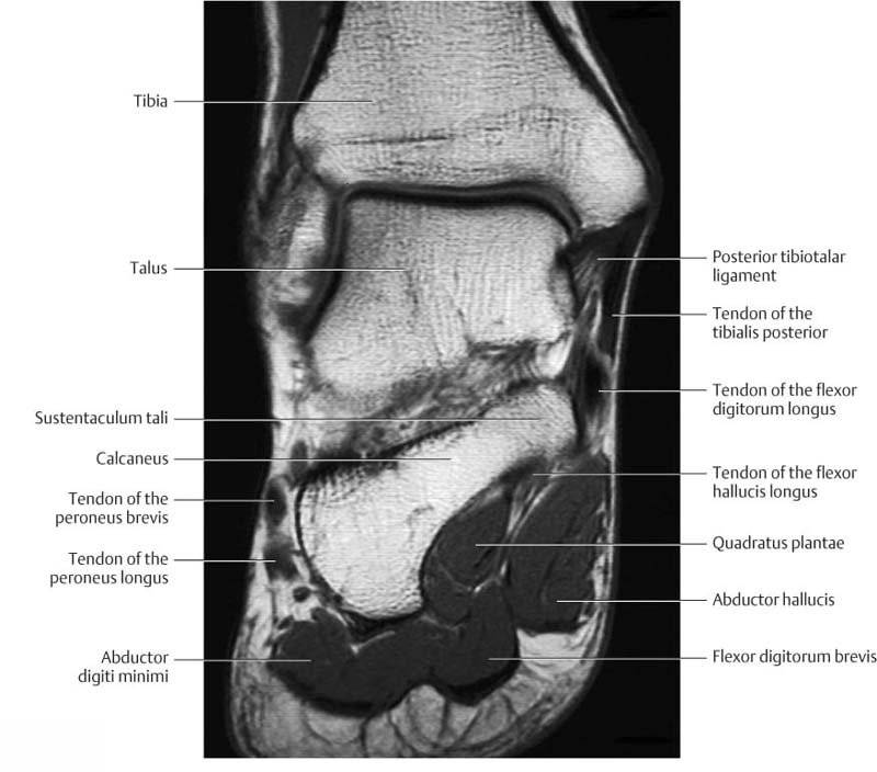
Anatomy of the foot and ankle - MRI from www.imaios.com Related posts of foot muscle anatomy mri muscle anatomy knee mri. Adductor hallucis is anatomically located in the central compartment of foot, but the muscle is functionally grouped with the medial plantar muscles of foot because it acts on the great toe (hallux). Magnetic resonance imaging, otherwise known as mri, uses a combination of magnetic fields and radio waves to take images of the internal structures of your body. Muscle anatomy knee mri 12 photos of the muscle anatomy knee mri muscle anatomy knee mri, human muscles, muscle anatomy knee mri Mri with user outlined plantar intrinsic and extrinsic muscles group. Chronic plantar fasciitis may be accompanied by muscle atrophy of plantar intrinsic foot muscles and tibialis posterior compromising the dynamic support of the foot prolonging the injury. Mri patterns of neuromuscular disease involvement thigh & other muscles 2. General anatomy and the musculoskeletal system:
Plantar interossei (foot) dr yuranga weerakkody ◉ and dr geon oh et al.
Magnetic resonance imaging (mri) is the modality of choice in diagnosing accessory muscles, delineating their relationship to adjacent structures, and differentiating them from soft tissue tumors. Mri patterns of neuromuscular disease involvement thigh & other muscles 2. General anatomy and the musculoskeletal system: They are mainly responsible for assisting some of the extrinsic muscles in their actions. Richard zimon answered 59 years experience internal medicine Anatomy of the whole human body : The paraspinal muscles, which are innervated by the spinal nerve dorsal ramus, are also frequently tested. During the first few days, this periosteal reaction may not be seen at conventional radiography because not enough calcium has. Related posts of foot muscle anatomy mri muscle anatomy knee mri. The interosseous muscles of the foot are muscles found near the metatarsal bones that help to control the toes. Mri is the choice of modality for further imaging the ankle and foot after obtaining initial radiographs. Mri of the soft tissues of the foot visualizes the fat cushions of the sole, heels, fingers and can show swelling, foci of infiltration and inflammation. Muscle anatomy knee mri 12 photos of the muscle anatomy knee mri muscle anatomy knee mri, human muscles, muscle anatomy knee mri